Beranda
/ Female Upper Back Anatomy - Hip Pain Explained Including Structures Anatomy Of The Hip And Pelvis / Other muscles are small and cover much less space.
Female Upper Back Anatomy - Hip Pain Explained Including Structures Anatomy Of The Hip And Pelvis / Other muscles are small and cover much less space.
Insurance Gas/Electricity Loans Mortgage Attorney Lawyer Donate Conference Call Degree Credit Treatment Software Classes Recovery Trading Rehab Hosting Transfer Cord Blood Claim compensation mesothelioma mesothelioma attorney Houston car accident lawyer moreno valley can you sue a doctor for wrong diagnosis doctorate in security top online doctoral programs in business educational leadership doctoral programs online car accident doctor atlanta car accident doctor atlanta accident attorney rancho Cucamonga truck accident attorney san Antonio ONLINE BUSINESS DEGREE PROGRAMS ACCREDITED online accredited psychology degree masters degree in human resources online public administration masters degree online bitcoin merchant account bitcoin merchant services compare car insurance auto insurance troy mi seo explanation digital marketing degree floridaseo company fitness showrooms stamfordct how to work more efficiently seowordpress tips meaning of seo what is an seo what does an seo do what seo stands for best seotips google seo advice seo steps, The secure cloud-based platform for smart service delivery. Safelink is used by legal, professional and financial services to protect sensitive information, accelerate business processes and increase productivity. Use Safelink to collaborate securely with clients, colleagues and external parties. Safelink has a menu of workspace types with advanced features for dispute resolution, running deals and customised client portal creation. All data is encrypted (at rest and in transit and you retain your own encryption keys. Our titan security framework ensures your data is secure and you even have the option to choose your own data location from Channel Islands, London (UK), Dublin (EU), Australia.
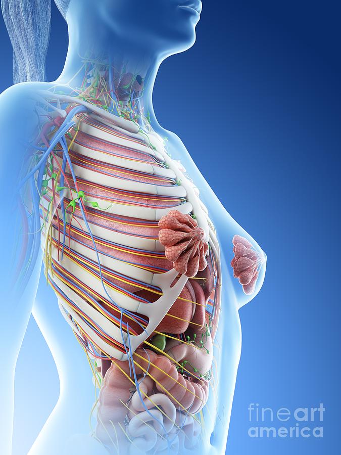
Female Upper Back Anatomy - Hip Pain Explained Including Structures Anatomy Of The Hip And Pelvis / Other muscles are small and cover much less space.. View of a dissected pumpkin female flower, or pistillate, with petals, corolla, stigma, style, ovary. Related posts of anatomy of the back organs human digestive system for kids. The bones of the chest and upper back combine to form the strong, protective rib cage around the vital thoracic organs such as the heart and lungs. Related posts of upper body of women antomy back women human anatomy. The back is found posteriorly and includes the vertebral column, the muscles that support the back and the spinal cord.
Understanding the anatomy of your lower spine can help you communicate more effectively with the medical professionals who treat your lower back pain. The cervical spine supports the weight and movement of your head and protects the nerves exiting your brain. The back's muscles start at the top of the back (named the cervical vertebrae) and go to the tailbone (also named the coccyx). Both the deltoid and the trapezius are firmly attached to … The cervical spine protects the nerves connecting to.
Female Upper Body Anatomy Photograph By Sebastian Kaulitzki Science Photo Library from images.fineartamerica.com The bones of the chest and upper back combine to form the strong, protective rib cage around the vital thoracic organs such as the heart and lungs. This muscle extends across the neck, shoulder, and back. 3d render of human body skeleton anatomy system. The trapezius and latissimus dorsi muscles connect the upper limb to the vertebral column. View of a dissected pumpkin female flower, or pistillate, with petals, corolla, stigma, style, ovary. The back is the body region between the neck and the gluteal regions. Female anatomy of internal organs with skeleton, rear and front views. It is the surface of the body opposite from the chest and the abdomen.the vertebral column runs the length of the back and creates a central area of recession.
The back anatomy includes the latissimus dorsi, trapezius, erector spinae, rhomboid, and the teres major.
The back functions are many, such as to house and protect the spinal cord, hold the body and head upright, and adjust the movements of the upper and lower limbs. Human body anatomy female female anatomy muscle shoulder blade pain anatomy back muscles bones man female anatomy body muscles in a body female anatomy muscole shoulder concept muscular sysyem. It is like that for several reasons, all of which you can understand by looking at the anatomy of the thoracic spine. The bones of the chest and upper back combine to form the strong, protective rib cage around the vital thoracic organs such as the heart and lungs. Related posts of upper body of women antomy back women human anatomy. The back anatomy includes the latissimus dorsi, trapezius, erector spinae, rhomboid, and the teres major. It runs from the neck to the upper back. Understanding the anatomy of your lower spine can help you communicate more effectively with the medical professionals who treat your lower back pain. On this page, you'll learn about each of these muscles, their locations and functional anatomy. 3d render of human body skeleton anatomy system. Each of the thoracic vertebrae are attached to the rib cage, providing a great deal of stability and structural support to protect the heart, lungs, and other important organs within the chest. The trapezius and latissimus dorsi muscles connect the upper limb to the vertebral column. Female student with a dissected cow brain.
3d render of human body skeleton anatomy system. Other muscles are small and cover much less space. Although it has many functions, the liver is best known for processing blood, separating waste from. The cervical spine supports the weight and movement of your head and protects the nerves exiting your brain. It is like that for several reasons, all of which you can understand by looking at the anatomy of the thoracic spine.
Female Anatomy Upper Back Ache Stock Photo Image Of Hair Human 34242346 from thumbs.dreamstime.com Related posts of anatomy of the back organs human digestive system for kids. Human digestive system for kids 8 photos of the human digestive system for kids circulatory system kids, human nervous system kids, human reproductive system kids, human respiratory system kids, human skeletal system kids, liver kids, pancreas kids, small intestine kids, human anatomy, circulatory system kids. Powerful muscles that move the head and arms attach to these bones as well. Female anatomy student, medical technologist or pathologist with a dissected cow brain slice through the mid section. The trapezius and latissimus dorsi muscles connect the upper limb to the vertebral column. It is the surface of the body opposite from the chest and the abdomen.the vertebral column runs the length of the back and creates a central area of recession. The breadth of the back is created by the shoulders at the top and the pelvis at the bottom. Learn to draw the upper back muscles by understanding the anatomical details and forms.
The upper back is called the thoracic spine, and it is the most stable part of the spine.
The vertebral column consists of 33 vertebrae which can be split up into 5 continuous sections. See human back anatomy stock video clips. This diagram depicts anatomy female 1024×1111 with parts and labels. Human body anatomy female female anatomy muscle shoulder blade pain anatomy back muscles bones man female anatomy body muscles in a body female anatomy muscole shoulder concept muscular sysyem. The cervical spine protects the nerves connecting to. Other muscles are small and cover much less space. The human back, also called the dorsum, is the large posterior area of the human body, rising from the top of the buttocks to the back of the neck. License image the deltoid, teres major, teres minor, infraspinatus, supraspinatus (not shown) and subscapularis muscles (not shown) all extend from the scapula to the humerus and act on the shoulder joint. Powerful muscles that move the head and arms attach to these bones as well. The major muscles in the upper torso of the body include: This diagram depicts anatomy female 1024×1111 with parts and labels. It is very stiff, and the thoracic spine has a limited range of motion. View of a dissected pumpkin female flower, or pistillate, with petals, corolla, stigma, style, ovary.
Although it has many functions, the liver is best known for processing blood, separating waste from. It is like that for several reasons, all of which you can understand by looking at the anatomy of the thoracic spine. Understanding the anatomy of your lower spine can help you communicate more effectively with the medical professionals who treat your lower back pain. The lumbar region of the spine, more commonly known as the lower back, is situated between the thoracic, or chest, region of the spine, and the sacrum. Some of these muscles are quite large and cover broad areas.
Back Muscles Chart By Badfish81 On Deviantart Muscle Diagram Back Muscles Human Back from i.pinimg.com The back is found posteriorly and includes the vertebral column, the muscles that support the back and the spinal cord. Female anatomy of internal organs with skeleton, rear and front views. Each of the thoracic vertebrae are attached to the rib cage, providing a great deal of stability and structural support to protect the heart, lungs, and other important organs within the chest. It comprises the vertebral column (spine) and two compartments of back muscles; This article looks at female body parts and their functions, and it provides an interactive diagram. The back anatomy includes the latissimus dorsi, trapezius, erector spinae, rhomboid, and the teres major. The rib cage also anchors the bones of the head, neck, shoulders, and arms to the trunk of the body. The 12 vertebrae in the upper back, labeled t1 down to t12, comprise the thoracic spine.
It runs from the neck to the upper back.
Related posts of anatomy of the back organs human digestive system for kids. Female anatomy of internal organs with skeleton, rear and front views. Function of the back muscles there are several individual muscles within the back anatomy, and it's important to take a quick look at all of The cervical spine is the top part of the spine. License image the deltoid, teres major, teres minor, infraspinatus, supraspinatus (not shown) and subscapularis muscles (not shown) all extend from the scapula to the humerus and act on the shoulder joint. Powerful muscles that move the head and arms attach to these bones as well. The range of motion in the upper back is limited because of the spine's attachments to the ribs (rib cage). The back's muscles start at the top of the back (named the cervical vertebrae) and go to the tailbone (also named the coccyx). Understanding the anatomy of your lower spine can help you communicate more effectively with the medical professionals who treat your lower back pain. The cervical spine supports the weight and movement of your head and protects the nerves exiting your brain. The 12 vertebrae in the upper back, labeled t1 down to t12, comprise the thoracic spine. The back anatomy includes the latissimus dorsi, trapezius, erector spinae, rhomboid, and the teres major. The lumbar region of the spine, more commonly known as the lower back, is situated between the thoracic, or chest, region of the spine, and the sacrum.
View of a dissected pumpkin female flower, or pistillate, with petals, corolla, stigma, style, ovary upper back anatomy. 3d render of human body skeleton anatomy system.